

When we think of fluorescence, highlighter pens, bright-white T-shirts and dresses glowing under nightclub lights, or SPF cream illuminating you like neon face paint in venues such as bowling alleys or crazy golf might come to mind. But there’s more to this phenomenon than meets the eye, and it’s contributing to observing everything from Alzheimer’s progression to cancer cell detection. Here, Knight Optical explains how fluorescence microscopes operate, their applications, and the optics that power them.

What is Fluorescence Microscopy?
Allowing users to see things like cell structures – which would otherwise be invisible to the naked eye – a fluorescence microscope highlights even the tiniest features in cells thanks to fluorescent indicators.
This is achieved by adding fluorophores – special dyes or fluorescent probes that fluoresce when exposed to selected wavelengths of light – to a specimen, such as a cell membrane, DNA or protein. The microscope then shines a light on the fluorophore, which absorbs energy and emits fluorescent light. With the support of optics in these instruments – such as excitation and emission filters – analysts can clearly see fluorescent substances against a dark background.
Different fluorophore variants glow in distinct colours. This means lab teams can choose particular fluorescent stains to trace various elements – for instance, often blue for DNA, red for mitochondria and fluorescent proteins like green fluorescent protein (GFP) for tagging targeted cell structures.
Real-World Uses
These fluorescent molecules can be applied to track changes as they happen or monitor gradual developments. As a result, this imaging approach is highly beneficial in many scientific fields. For example, in drug development, it can assist specialists in understanding how long it could take a pharmaceutical to reach its target; in disease diagnosis, it allows research teams to see how cancer cells metastasise; and in neuroscience studies, it can be adopted to evaluate cell-to-cell neural communication.
Fluorescence microscopy has changed the game for modern-day biology, making it possible for scientists to see proteins move, viruses invade cells and embryos grow – all in real time.

Different Types of Fluorescence Microscopy
While epifluorescence wide-field microscopy is the standard configuration for this technique, there are other, more advanced microscopes that give researchers the ability to see fluorescent proteins and probes with greater clarity and detail. Among them are confocal microscopes and super-resolution microscopes.
Confocal Microscopy
 Instead of a lamp, a laser powers the confocal microscope. Scanning point by point rather than lighting up the whole specimen, the laser in these devices produces crisp, high-contrast sections that are stacked into 3D visuals. With components like pinholes – which Knight Optical supplies in precision grades – blocking out-of-focus light, confocal systems are ideally suited to thicker samples.
Super-Resolution Microscopy
Perhaps the most celebrated form of super-resolution fluorescence microscopy is stimulated emission depletion (STED) microscopy. A Nobel Prize-winning breakthrough, STED microscopy overcomes diffraction limitations in conventional light microscopy by using a second doughnut-shaped laser – tuned to a precise STED wavelength – that switches off fluorophores around the outer edges. This leads to a resolution of 20-80nm, compared to approximately 250nm in confocal fluorescence microscopy.
Evanescent Wave Microscopy
Another approach implemented for its improved selectivity is total internal reflection fluorescence, also known as TIRF microscopy. Lighting up only around the first 100-200nm of a sample, TIRF microscopy generates clean, high-contrast images that allow users to view surface-level activity with almost no background noise.
Optical Components Used in Fluorescence Microscopes
At the core of every fluorescence microscope, a series of optics work together to maximise the signal-to-noise ratio, so users can obtain a clear image of their specimen and accurately assess movement and behaviour.
In the majority of configurations:
- Excitation filters narrow down the light source to the required wavelength for photon excitation of the fluorophore
- Dichroic mirrors/beamsplitters typically sit at 45º in the light path to reflect excitation wavelengths towards a sample while letting longer emission spectral ranges pass through to the detector
- Emission filters block out remaining excitation light and any stray signals to make sure only fluorescence reaches the eyepiece or camera in imaging systems.
At Knight Optical, we sell these components as fluorescence filter sets. These filter combinations are designed for specific fluorophores like CY5, ROX, TRITC, YFP, FITC and DAPI, and consist of a mounted excitation and emission filter with a 25mm diameter and a dichroic beamsplitter measuring 25.7mm x 36mm.
Why Optical Quality Matters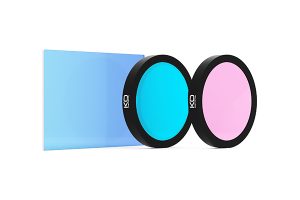
In cell biology and cell physiology research, optical quality plays a significant role in achieving clear results. Poor-quality filters can leak unwanted light, weak coatings degrade over time and inadequate components can blur the line between signal and noise.
When tracking protein-protein interactions, examining genetic material or imaging biopsied samples, there’s no room for error. High-quality optics not only ensure scientists obtain sharp, reliable images, but also help minimise sample damage by enabling efficient light transmission, thereby, reducing exposure time and reduced phototoxicity.
To achieve this standard of precision, our fluorescence filter sets are tested and quality-assured in our in-house metrology laboratory and QA department before dispatch, to confirm they meet the high expectations of fluorescence microscopy applications.
Whether you’re building a fluorescence setup or upgrading an existing system, get in touch with a member of our team today to find out how our precision optics can help.